Investigation
of the sub-cellular localization of Rqh1 in fission yeast
Okahira
Satoyo
Supervisor Dr. Elspeth Stewart, Dr. Miwa
Masanao
Background and Purpose
rqh1+ is the only member of the
RecQ helicase family found in Schizosaccharomyces pombe. There are other
homologues of RecQ helicases found in bacteria, yeasts, flies, frogs and
humans. Members of this DNA helicase family are interesting because they are
thought to be involved in maintaining genomic integrity. Mutation of members of
this family in humans is responsible for Werner's syndrome, Bloom's syndrome
and Rothmund-Thomson syndrome. Patients with these diseases show a
predisposition to cancer or premature ageing, and fission yeast cells with a
defective rqh1+ gene show reduced viability and abnormal
chromosomal loss even under normal growth condition.
To gain further insight into the role(s) of Rqh1 in S.
pombe, the sub-cellular localization has been investigated using indirect
immunofluorescence. Rqh1 is thought to be localized in the nucleus especially
in nucleolar foci. However this is not completely clear because of the
difficulty of seeing the nucleolus. Therefore nucleolar recognition was
attempted using two different methods to see where Rqh1 is localizing.
Before doing immunofluorescence, it was
necessary to purify a lot of anti-Rqh1 antibody, N156ECS. First
purification of the antibody was carried out using a modified Western blotting
method, called Western Method. However, this resulted in a lot of dilute
antibody which had to be concentrated x40 for immunofluorescence. For the
successful immunofluorescence, a rapid and high efficiency purification method
was constructed. It will be useful not only for the immunofluorescence study
but also for the other research into rqh1+
mechanisms.
Materials and Methods
The indirect immunofluorescence was done using Schizosaccharomyces
pombe, normal WT and rqh1¢ strains or WT and rqh1¢ leu- strains for
transformation. The anti-Rqh1 antibody, N156ECS, used in immunofluorescence was
purified from rabbit serum by using Western Method or Affi-Gel15 Method.
Affi-Gel15 Method was constructed using Affi-Gel15 agarose beads support
obtained from QIAGEN.
Result and Discussion
The new antibody purification method, the Affi-Gel15
Method, was successfully constructed. N156ECS purified by this method worked on
Western blotting and immunofluorescence and was x20- x40 more concentrated than
that purified using the Western Method. This method is also very useful
as there is no need to re-couple more antigen to beads to purify more N156ECS
once antigen is coupled to beads.
The plasmid containing GFP which localizes to the
nucleolus was transformed into yeast cells and cells showing good fluorescence
were obtained. Fixation conditions were revised for the use of these
transformed cells. Growing in YE5S medium instead of EMM Ahu medium is
necessary to fix the GFP transformed strains properly to use for
immunofluorescence. Rqh1 foci in normal WT cells were seen in the nucleolus.
And also in GFP transformed WT cells, Rqh1 looked like it was localizing to the
nucleolus but further experiments were required to confirm this.
Future work
Immunofluorescence examination using GFP transformed cells
should be done to confirm the localization of Rqh1 in the nucleolus. In
addition, using the delta-vision microscope is recommended to see the
fluorescence in more detail. It will then become possible to say exactly where
Rqh1 is localizing. Also it would be interesting to see whether the
localization of Rqh1 changes during each cell cycle or after DNA
damage.
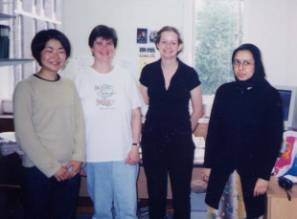
 I would like to thank Dr. Elspeth, my
supervisor, for her kindly help throughout my project (2nd from
left). And also, I would like to thank my co-worker, Fouzia (right) and
Caroline (2nd from right).